Diatoms are microscopic single-celled algae that have been present on Earth for at least 180 million years. They survive in any aquatic ecosystem with enough light - sea, lakes, rivers, even mud - where they live in the water mass as plankton or attached to plants, rocks or small sand particles. Diatoms are the most diverse protists on the planet (i.e. single-celled organisms with a nucleus), with around 20,000 known species, of which 90% - according to estimates - have not yet been discovered.
It is estimated that diatoms are responsible for 20% of total carbon fixation on Earth, being more productive than all the planet's tropical forests. They are therefore essential for the balance of the entire planet's ecosystem.
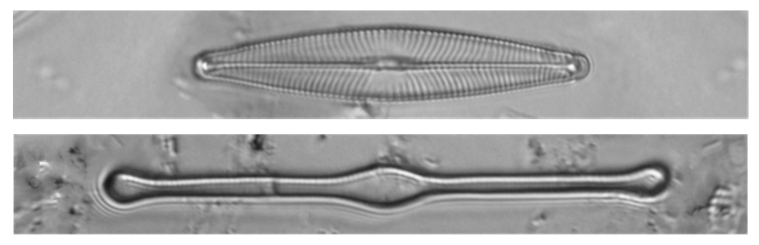
Due to the microscopic size of diatoms - ranging from 2μm to 2 mm in length for most species - they are invisible to the naked eye; therefore, scientists use optical microscopes or scanning electron microscopes (SEM) to reveal the secrets of diatoms. Diatoms are unique living beings whose cell walls are based on inorganic opaline silica, which is quite resistant to decomposition, heat and acids. It is as if the cell were inside a Petri dish, called a frustle. The cover consists of two intertwined halves, the epoxy and the mortgage - slightly smaller.
Each diatom species is adapted to a relatively specific ecosystem, so the quality of aquatic ecosystems can be inferred by obtaining indices of the diatom species present
They have the advantage that the silica skeleton of most types of diatoms is known to be tolerant to putrefaction, and that their collection in aquatic environments follows rapid and low-difficulty protocols. On the other hand, they have the disadvantage that the subsequent preparation of the samples in the laboratory is laborious and requires specialized instruments. In addition, to achieve their identification, the participation of expert taxonomists is required.
To mitigate this challenge, in this paper we propose a low-cost fully operational automated microscope, which integrates algorithms for (1) tape deck control and autofocus, (2) image acquisition (slide scanning, alignment, contrast enhancement), and (3) diatom detection and prospective sample classification (among 80 taxa). In-depth learning algorithms have been applied to overcome the difficult selection of image descriptors imposed by classical machine learning strategies. With respect to the mentioned strategies, the best results were obtained by deep neural networks with a maximum accuracy of 86% (with the YOLO network) for detection and 99.51% for classification, among 80 different species (with the AlexNet network). All the operating modules developed are integrated and can be controlled by the user from a developed graphical user interface running on the main controller. With the operating platform developed, it should be noted that this work provides a fairly useful set of tools in support tasks in the identification and classification of diatoms.
This work could give rise to technology transfer in the field of digital microscopy, incorporating future improvements to increase the system's capabilities and its possible adoption by the market. Some future work that could be carried out to improve the system would be
- Replacement of the small format PC by a computing unit with a GPU for embedded systems, capable of performing fast deep-learning inferences even for very demanding machine learning algorithms. In this line, a preliminary prototype is being tested with the NVIDIA Jetson platform.
-More accurate segmentation algorithms to reduce false positives and false negatives
-Improved modularity of mechanical elements, for rapid decoupling from standard microscopes.
-Faster response times for sequential scanning, applying more complex motor control strategies, minimising the effects of recoil and optimising the trajectories followed.
Link to the article
here