Wave-based optical coherence elastography: the 10-year perspective

Do you know what elastography is?
Summary of 10 years of research in elastography and future challenges to achieve the first commercial OCE elastograph
On the occasion of the 10th anniversary of the wave-based optical coherence elastography technique, two researchers from the Department of Biomedical Engineering from the University of Houston and the CSIC Institute of Optics have published a scientific article in which The development of the technique in these 10 years, its current state and its future challenges are summarized, with the aim of having the first wave-based OCE commercial device in the coming decades.
Optical coherence elastography based on mechanical waves is a very sophisticated technique that has become one of the main and most studied branches of elastography, which allows the exploration of the shape, size and hardness of internal tissues without the need to touch them.
This technique can be applied to soft (elastic) tissues such as the eyes, muscles, liver, brain, or skin to name just a few.
The oldest historical antecedent of elastography is palpation, in this way foreign elements (lumps) can be found inside the body because they are usually harder than healthy tissue. This technique is qualitative but not quantitative, it tells us if there is or not, but it does not serve to give precise measurements.
In turn, elastography measures the state of rigidity or elasticity of an internal tissue by sending mechanical waves (like a small blow) through them and detecting the shape and speed at which the vibration is transmitted in the different layers of the body. To “see” how this vibration is transmitted, other techniques capable of showing us the inside of the body are used, such as an ultrasound wave from ultrasound or a ray of light if the eye is being studied, so that it can be detected (through of the variations in the received signals) how the mechanical disturbance is transmitted inside the body.
In the case of the study of an eye, the mechanical disturbance can be a simple puff of air.

Two different types of materials being measured by elastography.
(a) design of new methods of excitation of mechanical waves in tissues,
(b) mathematical understanding of the propagation of mechanical waves in complex boundary conditions through the proposal of advanced analytical and numerical models,
and (c) the development of new sensors capable of recovering information from tissues in 2D and 3D.
These advances have allowed great advances in the improvement of the medical diagnosis of diseases and the monitoring of the treatment applied in various types of tissues, which makes the technique a firm candidate for clinical use in the future.
Beginnings of the technique
Historically, elastography was first implemented in imaging modalities such as magnetic resonance imaging (MRE) and ultrasound (USE) (with millimeter-scale image resolution) demonstrating extensive capabilities in generating 2D and 3D elasticity maps.
Later, optical coherence tomography (OCT) was developed, which uses infrared light to pass through the transparent parts of the eye and also different layers of the retina, reflecting part of each layer in order to provide information about the eye with a micrometric image resolution ( ∼3–10 μm) and a retinal penetration depth of 1 or 2 mm. This technique made possible the first implementation of elastography in OCT, also called optical coherence elastography (OCE).
This optical coherence tomography technology makes it possible to measure the vibration suffered by the tissues when receiving the mechanical wave with a sub-nanometric sensitivity, so that with a small external disturbance the system can be used to provide not only structural images of the internal tissues as in an ultrasound, but can detect its properties of elasticity or hardness.
Future challenges of elastography
The article explains that in order to improve the conclusions about the data received, it is necessary to understand even better the elastic properties of the tissues of the body, and in this area there is work to be done due to the complexity of some tissues, such as zones between two tissues without a well-defined boundary such as the corneoscleral boundary in the eye, effects of anisotropy, viscoelasticity like liver or non-linearity such as the increased stiffness of the cornea when it is stretched.
In addition, a lot of research is being done on the ways to send that wave that generates the elastic vibration of the tissues, there are those based on light, sound, air, electromagnetic or displacement pulses, there are those with contact or without contact such as blowing, and within them the vibration can be sent in many different ways to achieve different results or work better on different tissues or to avoid possible damage, infection and discomfort to the patient.
Related News
The Institute of Optics joins the celebration of 11F
Full and equal access and participation of women and girls in science and technology Madrid / January 31, 2025In recent decades, the international...
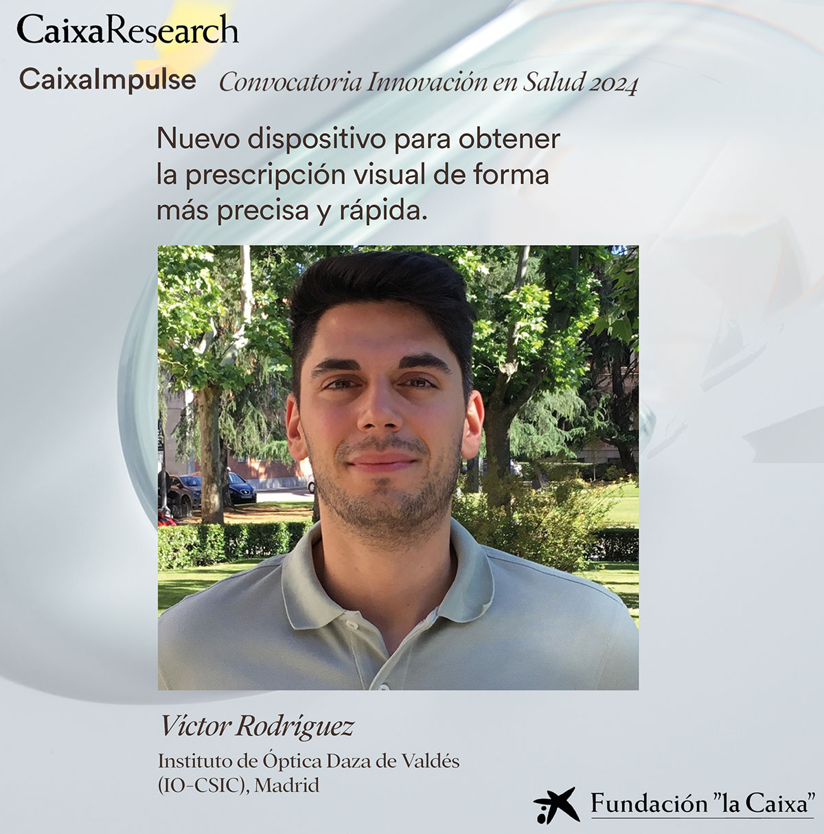
The SureVision research project has been awarded a Caixa Impulse innovation project, worth €500,000
Madrid / October 15, 2024Our colleague Víctor Rodríguez has managed to get his project SureVision to receive a €500,000 grant from the Caixa Impulse...

Predoctoral contract offer at the Laboratory of Visual Optics and Biophotonics
Madrid / October 2, 2024The Visual Optics and Biophotonics Laboratory has just published a job offer for a 4-year predoctoral contract within the...