Estimation of the full shape of the crystalline lens from OCT: validation using stretched donor lenses
-
The results demonstrate that this approach is accurate under both accommodation and misaccommodation conditions, making it promising for application in clinical practice.
Madrid / October 18, 2023
A major advance in the field of ophthalmology has been achieved by an international team of researchers and investigators, who in this work have validated their innovative method for estimating the complete shape of the human lens using coherence tomography images (OCT), and a prediction model for the shape of the lens called “eigenlenses”. This advance, published in the journal Biomedical Optics Express, has the potential to significantly improve treatments. of cataracts and presbyopia.
Últimas noticias

The lens of the eye is a crucial component that allows the focusing of near and far objects on the retina through the process of accommodation. However, with aging, the lens loses its transparency (cataracts) and hardens, losing its ability to accommodate (presbyopia). To improve current treatments for these conditions, it is essential to understand the geometry of the human lens and how it changes with age and accommodation.
In previous research, a model called “eigenlenses” was presented, which consists of a database of three-dimensional features that represent the complete shape of the ex vivo lens (the lens outside the eye). However, the challenge was to apply this model to estimate the full shape of the lens in vivo using OCT images, where only the central part of the lens can be visualized through the pupil.
In this new study, the researchers performed a validation of the use of “eigenlenses” to estimate the full lens shape in vivo under conditions of misalignment. They used 14 ex vivo donor lenses in a special setup and measured their geometry and power using a combined OCT and ray tracing aberrometry system. This allowed them to access the full extent of the lens and obtain measurements in both fully accommodated and unaccommodated conditions.
Subsequently, computational simulations were performed to recreate in vivo conditions. In these simulations it was assumed that only the central portion of the lens visible through the pupil was available. With these conditions and using the “eigenlenses” model, the complete shape of the misaligned lenses in vivo was estimated. The results showed a mean absolute error (MAE) between the estimated and measured lens diameters and volumes of MAE=0.26±0.18 mm and MAE=7.0±4.5 mm3, respectively.

The highlight of this study is that the estimation error did not depend on the accommodation state of the lenses, indicating that the “eigenlenses” method is equally accurate in both accommodation and misaccommodation conditions. This indicates that this approach can be used effectively to estimate the complete shape of misaligned lenses in vivo, which is of great relevance to improve current cataract and presbyopia treatments.
The results of this research open new possibilities for the development of more precise and personalized intraocular lens implantation treatments. By better understanding the geometry of the human lens, doctors will be able to make more accurate predictions of the power of the intraocular lens or the size of the lens to be implanted in a specific patient.
This is a collaborative research work of the Optics Institute of the CSIC, the Miami Bascom Palmer Eye Institute Ophthalmic Biophysics Center, Department of Engineering Biomedical, University of Miami, LV Prasad Eye Institute Ophthalmic Biophysics Laboratory of India, the Brien Holden Vision Institute of Sydney, the Department of Engineering of the Pontifical University of Peru and the Center for Visual Science of the University of Rochester, NY
IO-CSIC Communication
cultura.io@io.cfmac.csic.es
Related news
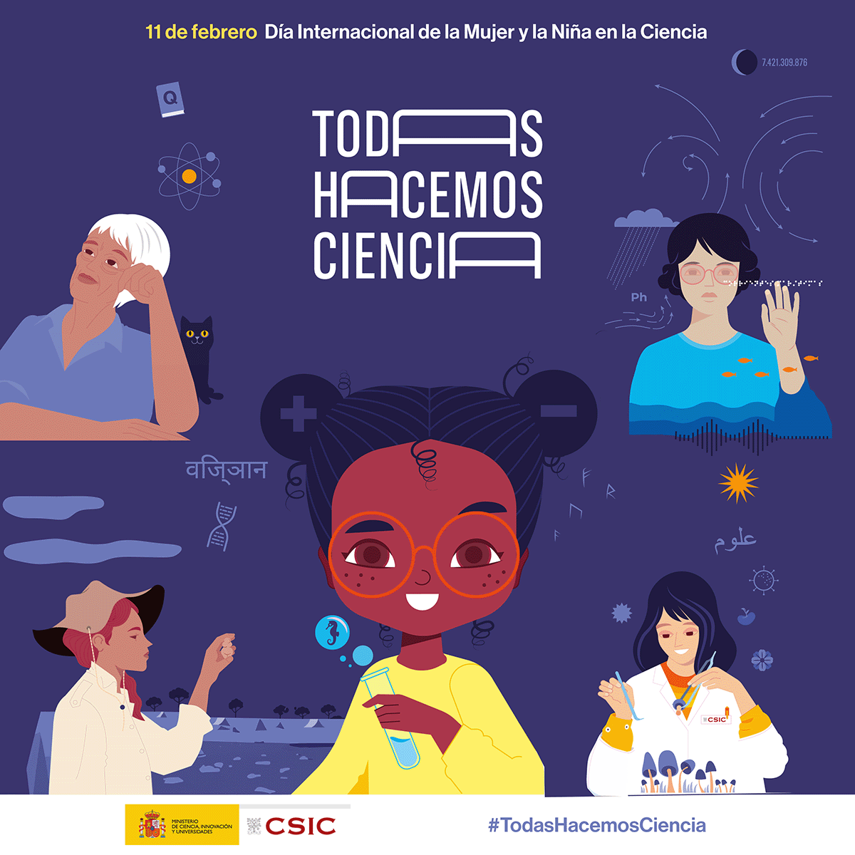
The Institute of Optics joins the celebration of 11F
Full and equal access and participation of women and girls in science and technology Madrid / January 31, 2025In recent decades, the international...
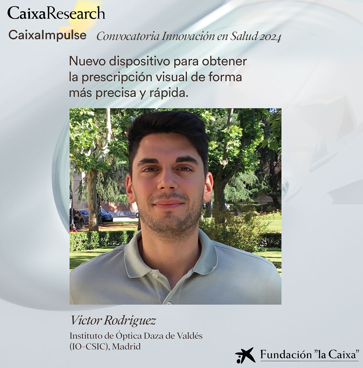
The SureVision research project has been awarded a Caixa Impulse innovation project, worth €500,000
Madrid / October 15, 2024Our colleague Víctor Rodríguez has managed to get his project SureVision to receive a €500,000 grant from the Caixa Impulse...
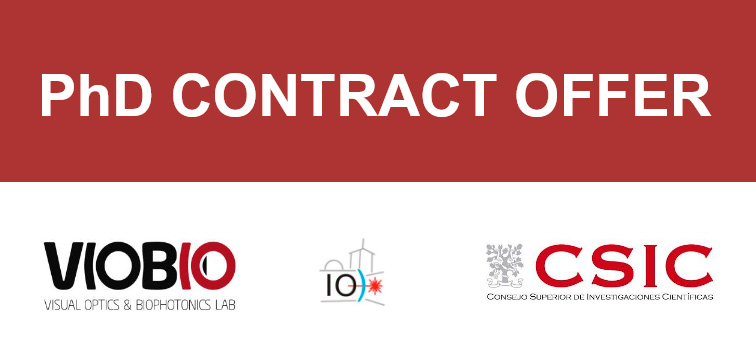
Predoctoral contract offer at the Laboratory of Visual Optics and Biophotonics
Madrid / October 2, 2024The Visual Optics and Biophotonics Laboratory has just published a job offer for a 4-year predoctoral contract within the...