A simple computational model for scleral stiffness assessments via air-puff deformation OCT
-
A new computational model allows noninvasive assessment of sclera stiffness, opening up new possibilities for diagnosing ocular pathologies.
-
The combination of air puff and recording of the resulting deformation by means of optical coherence tomography offers an advanced tool for studying the properties of the external structure of the eye in real time.
Madrid / September 10, 2024
Researchers from the Instituto de Óptica “Daza de Valdés” of the CSIC and the Center for Visual Science at the University of Rochester have published a study that presents a computational model to evaluate the stiffness of the sclera (the external, white part of the eye) through air puff deformation and OCT (optical coherence tomography) imaging. This advance offers a new tool to study the mechanical properties of the sclera in vivo, which could have significant implications for the diagnosis and treatment of pathologies such as myopia or keratoconus.
Latest news
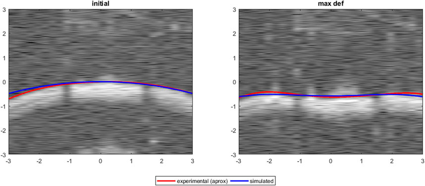
This study explores the use of a computational model that combines this technique with optical coherence tomography (OCT) to accurately and in real time assess sclera stiffness.
In this study, researchers developed a computational model using the finite element technique to simulate sclera deformation in response to an air puff. They used experimental data obtained from ex vivo rabbit eyes to validate the model. The model allowed parametric studies in which different parameters, such as tissue thickness, material properties, and intraocular pressure, were systematically varied to observe their influence on scleral deformation. The model results were compared to experimental measurements to assess its accuracy and ability to predict scleral stiffness, providing a robust tool to study the mechanical properties of the sclera noninvasively.

This study builds on previous research that has explored the use of corneal air-puff deformation imaging, extending and improving these techniques to assess scleral stiffness.
Article: Andres De La Hoz, Lupe Villegas, Susana Marcos, Judith S. Birkenfeld “A simple computational model for scleral stiffness assessments via air-puff deformation OCT”. Frontiers in Bioengineering and Biotechnology, 12, art. no. 1426060-esclerotica
IO-CSIC Communication
cultura.io@io.cfmac.csic.es
Related news
Thursday, February 29 scientific coffee with Víctor Rodríguez López
Madrid / February 12, 2024The following Scientific Café entitled "After 200 years: a new method of visual graduation" will be held. This talk will...
Diagnosis of ocular diseases by ocular biomechanics
Madrid / January 24, 2024Tomorrow, January 25, researcher Judith Birkenfeld will give a talk at the Fernández Vega Foundation.Judith Birkenfeld,...
SureVision research project, one of the 10 projects of technology companies supported by CSIC in 2023
Madrid / January 18, 2024The SureVision project of our colleague from the Visual Optics and Biophotonics Group, Víctor Rodríguez, has been one of...


